Video Laryngoscopes:
Structure, Purpose, and Evidence
Video laryngoscopes have established themselves as indispensable tools in anesthesia and emergency medicine in recent years. They allow for improved visualization of the laryngeal structures, thereby increasing the success rate of endotracheal intubation, especially in difficult airway conditions [1].
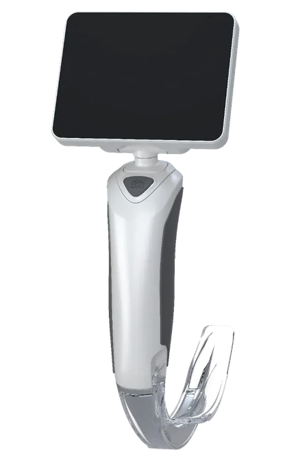
A video laryngoscope consists of several essential components:
Blade: It is inserted into the mouth to push the tongue to the side and provide a view of the glottis. There are different types of blades for various applications:
- Macintosh-like blade (allows direct and indirect visualization)
- Hyperangulated blade (suitable for difficult airway conditions) [2].
- Miller blade (suitable for use in neonates; see also here: Blade overview)
- Camera: A miniature camera attached to the tip of the blade transmits the image in real-time to a monitor [3].
- Light source: An LED light source provides optimal illumination of the airways [4].
- Monitor: The captured image is displayed on an external or integrated monitor, allowing for precise positioning of the endotracheal tube [5].
The main purpose of video laryngoscopes is the safe and efficient performance of endotracheal intubation. The most important areas of application include:
- Regular intubation: Especially in anesthesia, video laryngoscopes are increasingly used as a standard procedure [6].
- Difficult Airways: The S1 guideline for airway management recommends the primary use of video laryngoscopes in difficult intubation conditions [7].
- Emergency Medicine: Improved visibility of the airways can increase the success rate of emergency intubations [8].
- Medical Training: Due to the real-time display on a monitor, video laryngoscopes are ideal for training anesthetists and emergency medicine practitioners [9].
Scientific Evidence and Current Guidelines
According to the current S1 guideline "Airway Management" from the German Society for Anesthesiology and Intensive Care Medicine (DGAI) [7], a video laryngoscope should be available at every anesthesiology workstation. The main recommendations are:
- Video laryngoscopes with a Macintosh-like blade are suitable as the primary instrument for intubation [7].
- For anticipated difficult airways, hyperangulated blades should be available [10].
- After an unsuccessful direct laryngoscopy, the use of a video laryngoscope is recommended [7].
- Especially in patients at risk of aspiration, video laryngoscopy is the preferred method for securing the airway [11].
The introduction of videolaryngoscopes has significantly improved the clinical practice of airway management. Their benefits—particularly in difficult situations—are well substantiated by scientific studies and guidelines. Their routine use is increasingly regarded as the standard of care, and adequate training is essential to fully leverage the potential of this technology.
Video Laryngoscope
VX Series
Test our new models
VX-30 & VX-35.
Literaturverzeichnis
[1] Aziz, M. F., Healy, D., Kheterpal, S., et al. (2011). "Routine Clinical Practice Effectiveness of the Glidescope in Difficult Airway Management: An Analysis of 2,004 Glidescope Intubations, Complications, and Failures From a Prospective, Multicenter, Observational Study." Anesthesiology, 114(1), 34–41.
[2] Mosier, J. M., Sakles, J. C., Law, J. A., et al. (2020). "Tracheal Intubation in the Critically Ill: Current Status and Future Directions." The Lancet Respiratory Medicine, 8(8), 754–766.
[3] Cook, T. M., Kelly, F. E. (2017). "A National Survey of Videolaryngoscopy in the United Kingdom." Anaesthesia, 72(8), 1017–1024.
[4] Pieters, B. M., Maas, E., Knape, J. T. A., et al. (2017). "Videolaryngoscopy vs. Direct Laryngoscopy for Tracheal Intubation in Adults with Obesity: A Systematic Review and Meta-analysis." Anaesthesia, 72(6), 691–701.
[5] Myatra, S. N., Jain, R., Gandhi, K., et al. (2013). "Comparison of the C-MAC Videolaryngoscope with the Macintosh Laryngoscope for Routine Airway Management: A Randomised Clinical Study." Anaesthesia, 68(9), 899–907.
[6] Apfelbaum, J. L., Hagberg, C. A., Caplan, R. A., et al. (2013). "Practice Guidelines for Management of the Difficult Airway: An Updated Report by the American Society of Anesthesiologists Task Force on Management of the Difficult Airway." Anesthesiology, 118(2), 251–270.
[7] German Society of Anesthesiology and Intensive Care Medicine (DGAI). (2023). "S1 Guidelines for Airway Management." AWMF Registration Number: 001-028. Available at: https://register.awmf.org/assets/guidelines/001-028l_S1_Atemwegsmanagement_2023-09.pdf (accessed on February 4, 2025).
[8] Brown, C. A., Bair, A. E., Pallin, D. J., et al. (2010). "Techniques, Success, and Adverse Events of Emergency Department Adult Intubations." Annals of Emergency Medicine, 56(4), 260–270.
[9] Martin, L. D., Mhyre, J. M., Shanks, A. M., et al. (2011). "3,423 Emergency Tracheal Intubations at a University Hospital: Airway Outcomes and Complications." Anesthesiology, 114(1), 42–48.
[10] Piepho, T., Werner, C., Noppens, R. (2015). "Evaluation of the C-MAC Videolaryngoscope for Nasotracheal Intubation." Anaesthesia, 70(2), 134–138.
[11] Frerk, C., Mitchell, V. S., McNarry, A. F., et al. (2015). "Difficult Airway Society 2015 Guidelines for Management of Unanticipated Difficult Intubation in Adults." British Journal of Anaesthesia, 115(6), 827–848.